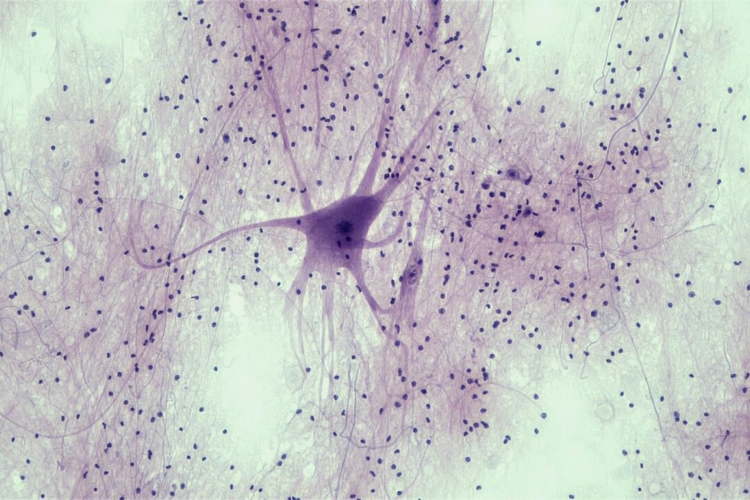
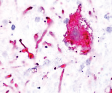
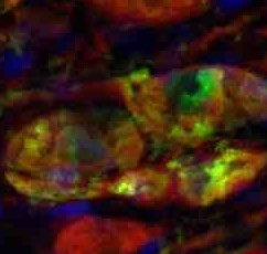
α-Synuclein is the major constituent protein of aggregates in Lewy bodies, the pathological hallmark of Parkinson’s disease. Viral delivery of α-synuclein to the substantia nigra of non-human primates (Macaca fascicularis) produces a slowly developing degeneration of dopaminergic neurons in the substantia nigra with pathology similar to that observed in people with Parkinson’s disease.
- Disease-relevant mechanism of dopaminergic degeneration
- Imaging allows longitudinal monitoring of dopaminergic neuron function
- Slowly developing primate model of synucleinopathy
Model Overview
AAV1/2-A53T-α-synuclein vectors are injected bilaterally into the substantia nigra of macaques using MRI-based stereotactic techniques. Two injection sites per hemisphere are used to ensure complete viral coverage of the substantia nigra. Degeneration of dopaminergic neurons occurs over several weeks allowing test compounds to be evaluated for disease modifying potential. Some implementations of the model provide behavioural and imaging deficits, in addition to pathology readouts.
Robust loss on dopaminergic endpoints
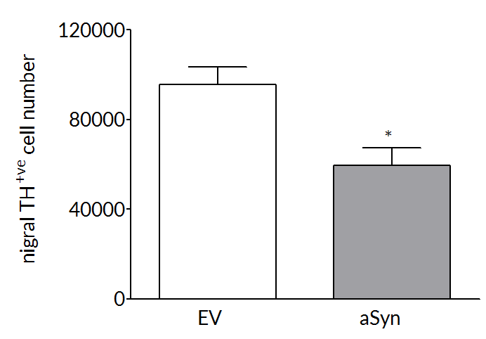
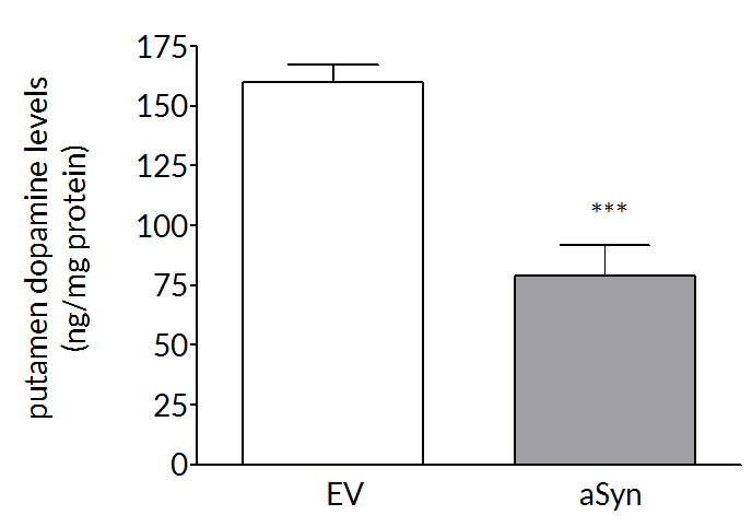
Dopaminergic cell loss develops over time so that at 17 weeks post-surgery, there is a 40-50% loss of striatal dopamine and tyrosine hydroxylase positive cells in the substantia nigra.
Test compounds can reverse AAV αSyn induced loss of dopamine
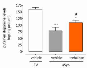
The loss of striatal dopamine that occurs over 17 weeks post-surgery can be significantly attenuated by daily administration of trehalose, a known autophagy enhancer, demonstrating that compounds that have the potential to slow disease progression can be evaluated in this model.
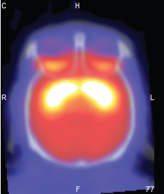
PET imaging allows in-life assessment of dopaminergic cell loss
Striatal levels of markers of the dopaminergic system (DAT and VMAT) can be imaged throughout longitudinal studies – allows the effect of compounds to be followed over time.
Experimental readouts
- Behavioural – Behavioural assessments include the monkey parkinsonian disability rating scale, the monkey equivalent of the clinical rating scales used to assess in people with Parkinson’s disease. Additional behavioural endpoints include home-cage activity, observation-cage activity and fine motor control.
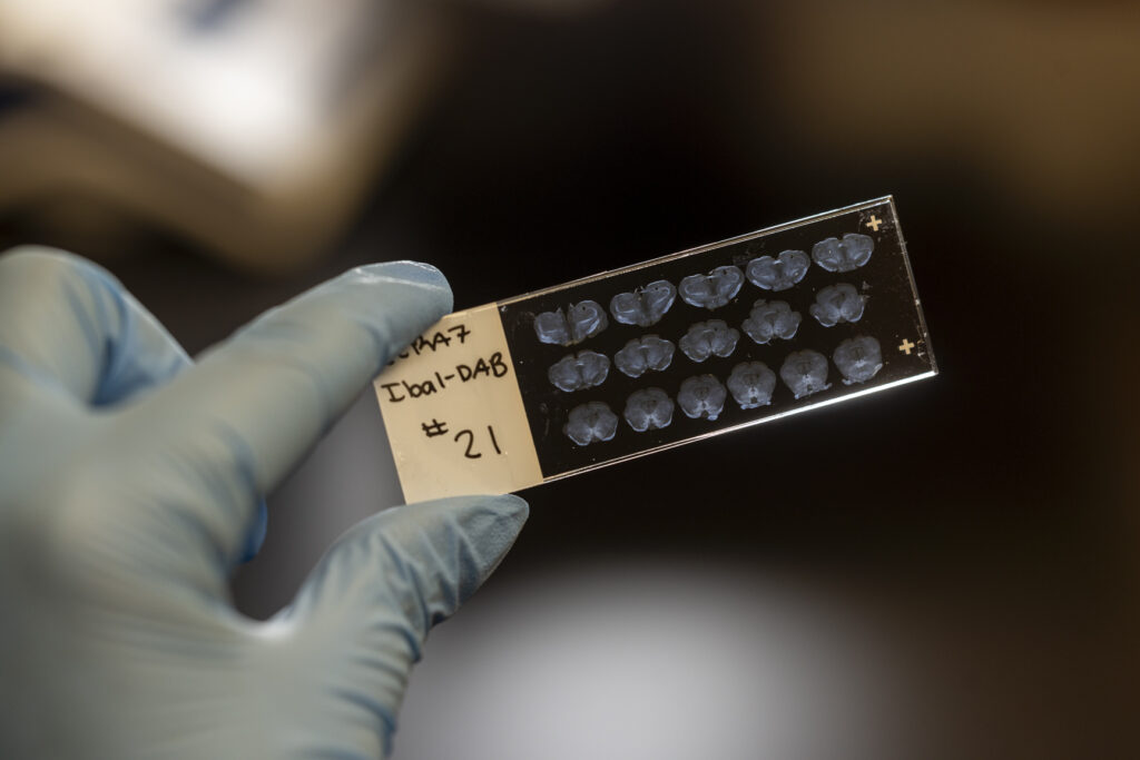
- Post mortem – Routine post-mortem analyses include striatal dopamine and dopamine transporter (DAT) and the number of number of TH+ve cells in the substantia nigra. Additional post-mortem measures can be incorporated at the request of the client.
- Imaging – We offer both MRI and PET imaging that allows longitudinal measurement of markers of dopaminergic function and metabolism.
- Pharmacokinetics, safety and blood chemistry – Can be incorporated into all studies. Blood and CSF can be sampled throughout the study and terminal samples of brain and other tissues can be collected. Functional observational battery and blood chemistry can be used to assess off target and adverse effects.